Single cells and the master clock
In mammals, the suprachiasmatic nucleus (SCN) in the hypothalamus is the central pacemaker of daily rhythms in sleep, body temperature and hormones. Its neurons stay tightly synchronized.
Overview
The Granada Lab (2020–2025) was a computational biology group in biomedical research at the Comprehensive Cancer Center of Charité University Hospital Berlin. We addressed these questions with precision-medicine approaches that combined laboratory experiments, computation and mathematical modelling, in collaboration with clinical oncologists.
We were committed to training students and young researchers, and to making our data and software reproducible, reliable and openly available.
Approach
Long-term live-cell imaging. We set up an S2-certified live-cell imaging facility and a high-throughput deep-phenotyping pipeline that records clock and growth dynamics in single cells across hundreds of conditions.
Computational biology. Time-series analysis and mathematical models turn these recordings into interpretable features and testable predictions. The group was the first computational biology unit within the Comprehensive Cancer Center at Charité.
Models before animals. Models guide the experiments, predict outcomes and reduce the need for animal testing.
Toward patients. Since 2025 these methods have moved toward application: a treatment-optimization platform in the BIH Digital Health Accelerator, and an article for oncologists on the timing of cancer therapy.
Research focus · Treatment timing
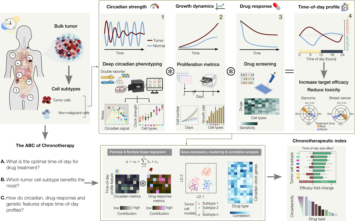
The circadian clock regulates many cellular processes, and its disruption, as in shift work, has been linked to cancer risk. Using circadian rhythms to time treatment is promising but still emerging. We built a framework to make it measurable.
With a high-throughput deep-phenotyping pipeline we showed that the time of administration alters the effect of a broad panel of cancer drugs. Circadian features differ enough between cells to define subtypes with distinct drug sensitivities: first in breast cancer, where four clock phenotypes emerged, and then in neuroblastoma, where clock heterogeneity accounts for part of the variability in response. Together, these results turn time-of-day effects from an observation into a cell property that can be assayed, classified and, in principle, acted on.
Research focus · Surviving therapy
After chemo- or radiotherapy, some tumor cells die, some stop dividing and some survive and keep proliferating. This fractional response is one reason treatment fails: a subpopulation survives, while patients still bear the side effects.
In earlier work at Harvard Medical School with Galit Lahav, we showed that survival under chemotherapy is set by a cell’s proliferative history and cell-cycle position. In the lab we showed that the joint dynamics of p53 and p21 encode how much DNA damage a cell sustained, fine-tuning whether it proliferates and shaping heterogeneity across the population; and, with collaborators, that inheritable cell states, not chance alone, determine which cells persist under drug treatment.
Single-cell state-space trajectories of three internal variables (circadian clock, cell cycle, p53):
Cell #2
Cell #3
Cell #4
Research focus · Clock and cell growth
Over fifteen years we developed a quantitative account of how circadian oscillators synchronize. Coupling strength and oscillator amplitude set the range of cycles a clock can follow: strongly coupled clocks are more robust but harder to shift, which explains why tissues re-entrain at different speeds. The prediction was later confirmed when coupling between peripheral oscillators was shown to be carried by TGF-β signaling.
We then showed what this coupling is for: it orchestrates cell growth, locking the circadian oscillator to the cell cycle, with coupling strength, not the mere presence of a clock, determining the outcome. This also reconciles reports that circadian disruption can both promote and suppress tumor growth: the sign of the effect depends on the properties of the cells involved.
Coupled U2OS cells
Uncoupled U2OS cells
Research focus · Methods for others
Single-cell rhythms are noisy and non-stationary, and their analysis is easy to get wrong. We made our methods available: pyBOAT, open-source software for time-frequency analysis of biological oscillations, and an accessible guide to wavelet analysis of circadian data. Code is openly available on GitHub; tutorials and recordings are on the Resources page.
Before 2020
Earlier work, with the labs of Hanspeter Herzel (Humboldt University Berlin) and Achim Kramer (Charité), that underpins our approach to treatment timing.
In mammals, the suprachiasmatic nucleus (SCN) in the hypothalamus is the central pacemaker of daily rhythms in sleep, body temperature and hormones. Its neurons stay tightly synchronized.
SCN and peripheral tissues share almost identical molecular clocks but respond very differently to periodic signals. Combining theory with whole-tissue and single-cell experiments, we showed that intercellular coupling explains these differences, identified what sets the speed of entrainment, and modelled how the SCN network can split into two rhythms.
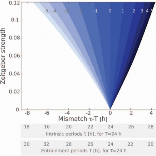
Why are some people “early birds” and others “night owls”? We provided a framework explaining the distribution of human chronotypes and how wake-up time depends on season and on clock properties such as relaxation time and intercellular coupling.
Phase response curves quantify how oscillators respond to pulse-like perturbations. Combining them with information theory, we studied temperature cycles as a time cue for peripheral clocks and tested the predictions experimentally.
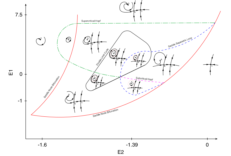
For my master’s thesis, with Marcos Trevisan and Gabriel Mindlin at the Dynamical Systems Lab in Buenos Aires, I used bifurcation theory and simulations to study the respiratory rhythms that drive birdsong.